Landmark clinical evidence for intraoperative breast margin assessment in breast-conserving surgery
Published in JAMA Surgery, the BrIMA study demonstrates how high-resolution specimen PET-CT imaging can support more informed intraoperative margin management during breast-conserving surgery.
Download the study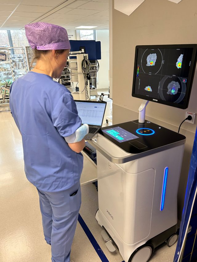
Positive margins continue to challenge breast-conserving surgery
Despite advances in breast-conserving surgery, positive margins remain a common and clinically significant challenge. Conventional intraoperative assessment may not identify all positive margins, potentially resulting in additional surgery, increased patient burden, and higher healthcare costs.
In the BrIMA study, almost 1 in 4 patients had a positive margin after the initial breast tumor resection — in line with positive margin rates reported in the breast-conserving surgery literature.
Because definitive histopathology is not available during surgery, surgeons must often make margin-management decisions with incomplete information. The BrIMA study evaluated whether intraoperative specimen PET-CT imaging could improve identification of positive margins while the patient remained in the operating room.
patients had a positive margin after the initial tumor resection
Prospective, multicenter clinical evidence
BrIMA is the largest prospective, multicenter clinical study evaluating high-resolution specimen PET-CT imaging for intraoperative breast margin assessment in breast-conserving surgery. Conducted across six European breast cancer centers, the study compared specimen PET-CT imaging with standard intraoperative assessment, using permanent histopathology as the reference standard.
Primary endpoint achieved
Intraoperative margin assessment success rates were numerically higher with AURA 10® PET-CT than with routine assessment alone.
Intraoperative success rate with specimen PET-CT imaging in the IDC group.
PET-CT findings altered intraoperative management in more than 1 in 6 patients.
Good interobserver agreement.
PET-CT achieved the highest intraoperative success rate for addressing invasive positive margins during breast-conserving surgery.
Specimen PET-CT reached a 95.2% intraoperative success rate in the IDC group, compared with 83.3% for no intraoperative margin assessment (p < 0.001) and 86.9% for standard of care (p = 0.327).
By providing additional information during surgery, AURA 10 helps surgeons identify positive margins that might otherwise only be discovered after postoperative pathology review, allowing corrective action while the patient remains in the operating room.
Ghent University Hospital, Belgium
What BrIMA means in clinical practice
Supports more informed intraoperative specimen evaluation.
Provides additional information before surgery is completed.
Integrates into existing breast-conserving surgery workflows with a 10-minute specimen scan.
Immediate specimen-level insight at the point of surgery
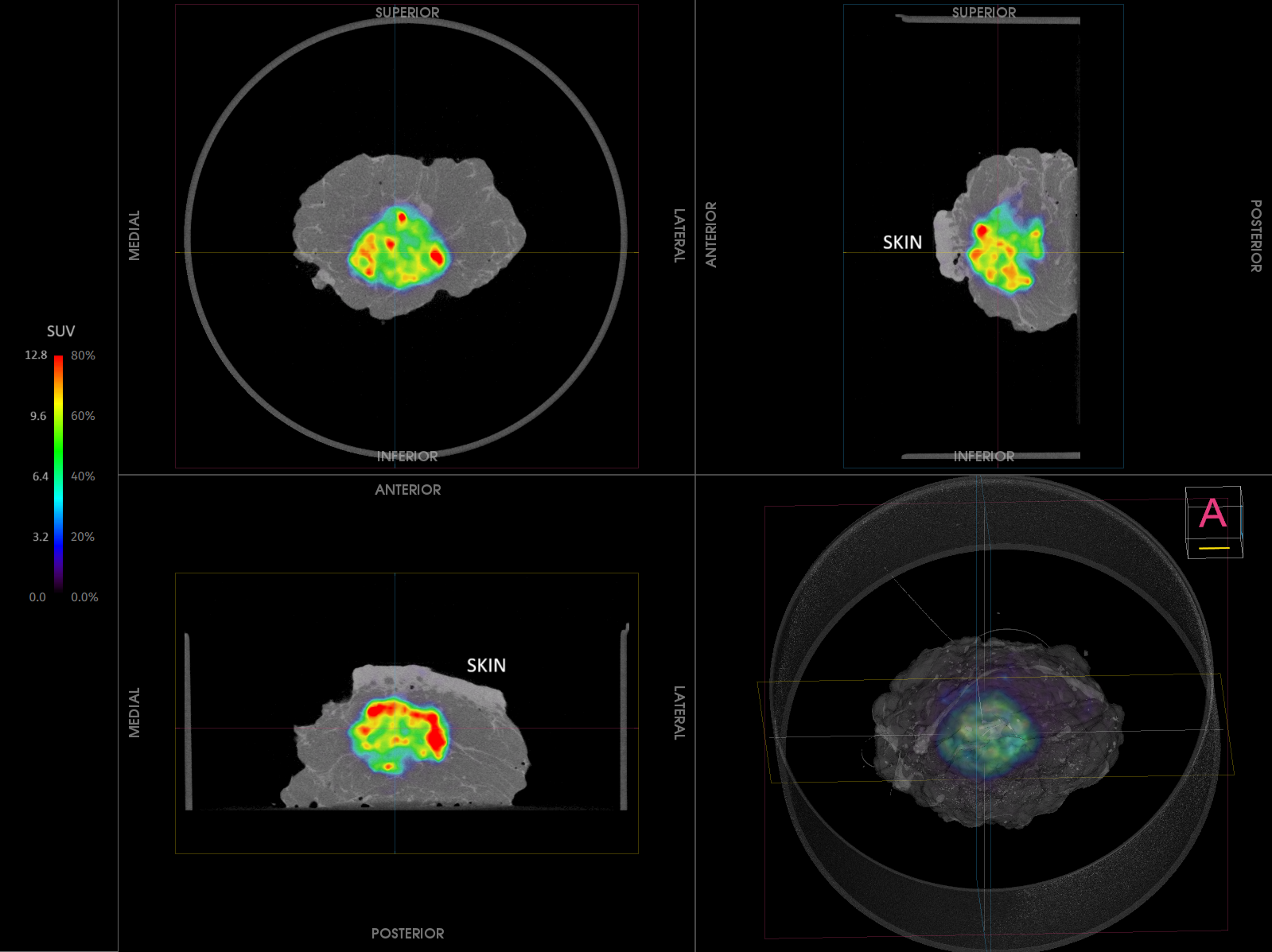
AURA 10® combines high-resolution PET and CT imaging to provide three-dimensional visualization of the surgical specimen during breast-conserving surgery.
Designed for use within the surgical workflow, AURA 10 provides additional intraoperative information while supporting intraoperative assessment and decision-making.

Read the complete JAMA Surgery publication and explore the clinical evidence supporting specimen PET-CT imaging during breast-conserving surgery.
Advancing the evidence for intraoperative imaging
BrIMA represents an important milestone in the growing body of clinical evidence supporting specimen PET-CT imaging as an adjunct to conventional intraoperative assessment.