Imaging Case 22:
Prostate cancer - Adenocarcinoma
Radical prostatectomy is a surgical procedure that aims to remove the entire prostate gland and, in many cases, the surrounding tissues, including the seminal vesicles and nearby lymph nodes. It is a primary treatment option for localized prostate cancer and aims to achieve long-term cancer control while preserving urinary and sexual function whenever possible.
A key objective of radical prostatectomy is to achieve negative surgical margins —meaning that no cancer cells are found at the edges of the removed tissue. This indicates that the tumor has been fully excised, reducing the risk of local recurrence and improving long-term outcomes. More specifically, patients with negative margins are less likely to require adjuvant therapy, which can cause additional side effects such as fatigue, urinary incontinence, and erectile dysfunction.
Intraoperative information may play a valuable role in ensuring the completeness of resection. Recent studies have demonstrated that high-resolution specimen PET-CT imaging using PSMA-based radiotracers provides a detailed view of the resected prostate tumor, potentially aiding in the assessment of surgical margins and enhancing surgical precision [1,3,4,5].
In this case, we illustrate how intraoperative specimen PET-CT imaging with 18F-PSMA can visualize the tumor within the resected tissue specimen. When combined with all available diagnostic data, the specimen PET-CT images can support the surgical team in assessing whether additional tissue removal is necessary to optimize surgical outcomes.
This case is presented with the support ofAZ Maria Middelares, Ghent, Belgium, and is part of the investigator driven study to explore potential indications of an intraoperative specimen PET-CT imager (Trial registration number: BUN: B0172022000009).
Patient history
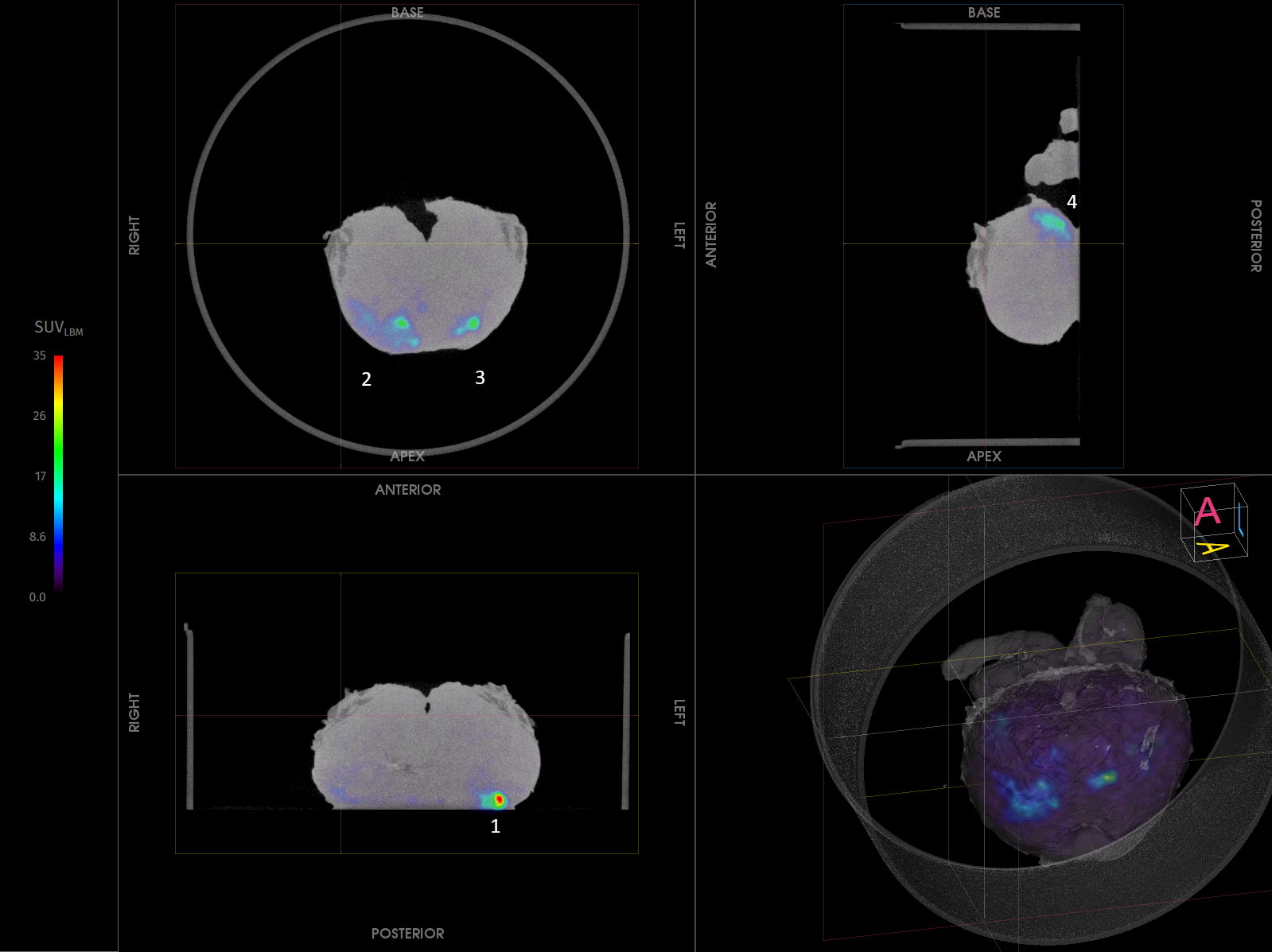
A 80-year-old patient was diagnosed with invasive prostatic adenocarcinoma (Gleason score 4+3). Preoperative imaging, i.e., MRI and 18F-PSMA PET-CT, showed several lesions at the left, right and anterior side of the prostate. Biopsy confirmed from both the right and left sides moderately differentiated invasive prostate adenocarcinoma, with a Gleason score of 4 + 3. The patient was staged as cT2cN0M0 and scheduled for robot assisted prostatectomy.Figure 1. Transverse, coronal, and sagittal slices and 3D view of the specimen PET-CT images of the prostate specimen. Specimen orientation is as indicated. The tumor is represented by a bright colorful region in the specimen. The PET tracer scale bar is depicted on the right-hand side. At the left posterior zone (1) of the specimen, 18F-PSMA uptake is seen at the border of the specimen.Specimen PET-CT imaging
The patient was intravenously injected with 1 MBq/kg of 18F-PSMA during surgery.
Resection of the prostate specimen was completed at 168 min after injection. Immediately after resection, a high-resolution specimen PET-CT image was acquired in the operating theatre. The images are shown in Fig. 1. In this image three orthogonal views of the specimen PET-CT images are shown. The PET images are represented in color scale, superimposed on the CT images in greyscale. The images of the prostate specimen clearly show increased 18F-PSMA uptake. Four clear 18F-PSMA uptake regions are seen, for which the PET uptake at the left posterior zone (1) that reaches the border of the specimen, see Fig. 1.
Histopathological evaluation
After specimen PET-CT imaging, the surgical specimen was sent to the pathology department for routine histopathological evaluation, which was available after several days. The histopathological results are listed in Table 1.
A positive surgical margin was found at the left posterior border of the specimen. This correlates with the region of radiotracer uptake on the specimen PET-CT image that reaches the tissue border. Additionally, all lesions seen on specimen PET-CT imaging with radiotracer uptake at a distance of the specimen border, were also seen on histopathology with a negative margin.
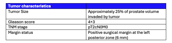 Table 1. Tumor characteristics assessed by histopathological evaluation.
Discussion and conclusion
A primary goal of radical prostatectomy is achieving negative surgical margins, ensuring that no cancer cells remain at the edges of the removed tissue. This significantly reduces the risk of local recurrence and improves long-term patient outcomes. Patients with negative margins are less likely to need adjuvant therapy, which can lead to fatigue, urinary incontinence, and erectile dysfunction.
In this case, specimen PET-CT imaging revealed 18F-PSMA uptake in four regions, with uptake at the posterior left side reaching the specimen border. These findings correlated with preoperative MRI and PET-CT and were confirmed by histopathology, which identified these lesions as tumors, including a positive surgical margin at the left posterior border.
By providing real-time insights, intraoperative specimen PET-CT gives surgeons a clearer picture at the point of surgery, which may enable more precise resections and potentially reduce the need for adjuvant therapy. This approach aims to enhance oncological outcomes, minimize complications, and improve quality of life for patients.
References
[1] Darr C et al. (2023). Intraoperative Molecular Positron Emission Tomography Imaging for Intraoperative Assessment of Radical Prostatectomy Specimens. European Urology Open Science 54:28-32
[2] Okarvi, S. M. et al. (2019). Recent developments of prostate-specific membrane antigen (PSMA)-specific radiopharmaceuticals for precise imaging and therapy of prostate cancer: an overview. Clin. Transl. Imaging 7:189–208. issn: 22817565
[3] Muraglia L et al. (2023). First Live-Experience Session with PET/CT Specimen Imager: A Pilot Analysis in Prostate Cancer and Neuroendocrine Tumor. Biomedicines 11:645.
[4] Lambert B. et al. (2025) Feasibility study on the implementation of a mobile high-resolution PET/CT scanner for surgical specimens: exploring clinical applications and practical considerations. Eur J Nucl Med Mol Imaging. https://doi.org/10.1007/s00259-025-07143-z
[5] Moraitis A. et al. (2025) Evaluation of Surgical Margins with Intraoperative PSMA PET/CT and Their Prognostic Value in Radical Prostatectomy. jnumed. 124.268719. https://doi.org/10.2967/jnumed.124.268719