Head & Neck Cancer - Oral Tongue Squamous Cell Carcinoma
Achieving adequate surgical margins is a critical determinant of oncologic outcomes in head and neck cancer. In anatomically complex regions such as the oral tongue, complete tumor resection must be balanced with preservation of function, making intraoperative decision-making particularly challenging. Close or involved margins may necessitate additional treatment, including re-resection or adjuvant therapy, both of which can significantly impact patient morbidity and quality of life [1].
Intraoperative specimen PET-CT imaging has emerged as a technique that may provide real-time insight into tumor extent and margin status, thereby supporting surgical assessment during resection [2].
This case is presented with the support of Dr. Michael C. Topf, Associate Professor, Head & Neck Surgical Oncology – Microvascular Reconstruction, Department of Otolaryngology, Vanderbilt University Medical Center, Nashville, TN, United States.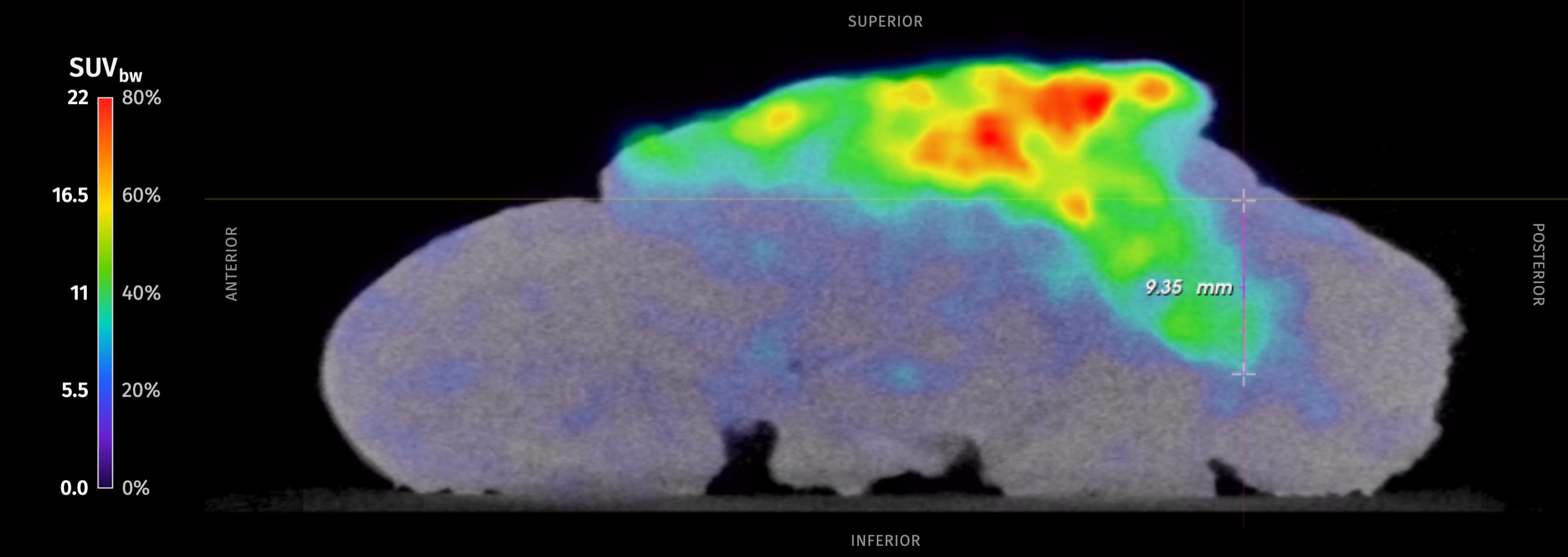
Patient History
A 55-year-old patient presented with a lesion of the anterior midline of the oral tongue. A biopsy showed at least squamous cell carcinoma in situ. Preoperative CT showed no regional nodal or metastatic disease.
Given these findings, the patient was scheduled for a partial glossectomy. Neck dissection was delayed as depth of invasion and need for reconstruction were unclear preoperatively.
Surgical Procedure
The procedure was performed at Vanderbilt University Medical Center by a surgical team led by Dr. Topf. The patient underwent an anterior glossectomy that removed the anterior third of the oral tongue, followed by partial primary closure. The tumor was exophytic, broad-based and about 3.5 cm in size, just right of the midline. It had a deeper component just right of the midline posteriorly. The floor of the mouth was clear and was preserved bilaterally.
Because a third of the tongue had been removed, the surgeon delayed the neck dissection. The plan was to assess the patient's function over the following weeks and decide then whether free flap reconstruction was needed, at the time of the elective neck dissection.
Figure 1: High-resolution PET-CT image from the anterior oral tongue, sagittal view. The tumor’s depth of invasion was measured as 9 mm. SUVmax (BW) = 27.Specimen PET-CT Imaging
The patient received an intraoperative injection of 0.8 MBq/kg (0.022 mCi/kg) of 18F-FDG per study protocol. Approximately one hour after injection, the primary tumor specimen was excised and imaged with the AURA 10 PET-CT.
The intraoperative specimen PET-CT images demonstrated radiotracer uptake within the specimen corresponding to the tumor, which did not extend to the posterior, anterior, lateral, medial and deep margins. The tumor’s depth of invasion was estimated at 9 mm on the intraoperative PET-CT images. No additional resection was performed based on the intraoperative imaging and gross examination.
Histopathological Evaluation
Final histopathological evaluation confirmed a unifocal, invasive, keratinizing squamous cell carcinoma, moderately differentiated (G2). It was located in the midline of the anterior two-thirds of the tongue and measured 3.5 cm in greatest dimension. The depth of invasion was 9 mm, with no lymphovascular or perineural invasion identified. The tumor was classified as pT2.
All margins of the specimen were negative for invasive tumor and for high-grade dysplasia or in situ disease. The closest margin was the inferior/deep soft tissue margin, at 4.5 mm. The other margins measured 14 mm (posterior mucosal), 13 mm (right lateral) and 8 mm (left lateral).
Discussion and Conclusion
This case shows the value of intraoperative specimen PET-CT imaging during an anterior glossectomy for oral tongue squamous cell carcinoma. The tumor’s depth of invasion was a key open question going into surgery, because the decision to perform elective neck dissection depended on it.
The depth of invasion estimated on the specimen PET-CT images was 9 mm, the same value reported on final histopathology. The images also showed the tumor clearly within the resected specimen.
All relevant resection margins showed no radiotracer uptake extending to the specimen border, suggesting that no invasive tumor reached the margins. These findings matched final histopathology for every margin.
This case highlights the potential of depth of invasion on specimen PET-CT images during surgery to inform margin assessment and decisions on neck dissection. Used in real time, this information could support the surgeon's assessment of resection completeness and the necessity for neck dissection.
References
- Machiels JP, Leemans CR, Golusinski W, et al. Squamous cell carcinoma of the oral cavity, larynx, oropharynx and hypopharynx: EHNS–ESMO–ESTRO Clinical Practice Guidelines. Ann Oncol. 2020;31(11):1462–1475.
- Lambert B. et al. (2025) Feasibility study on the implementation of a mobile high-resolution PET/CT scanner for surgical specimens. Eur J Nucl Med Mol Imaging. https://doi.org/10.1007/s00259-025-07143-z