Imaging Case 38
Head & Neck Cancer - Metastatic Squamous Cell Carcinoma of the Neck, Unknown Primary
In patients with head and neck squamous cell carcinoma, management of the neck is essential to treatment [1]. Accurate assessment of metastatic nodal involvement is therefore important, and lymph nodes that remain indeterminate on preoperative imaging can complicate surgical decision-making.
High-resolution specimen PET-CT imaging with 18F-FDG has been shown to be feasible for the three-dimensional assessment of resected head and neck cancer specimens in the operating room [2, 3]. It may also provide immediate insight into radiotracer uptake in lymph nodes to support assessment during surgery.This is presented with the support of Dr. Michael C. Topf, Associate Professor, Head & Neck Surgical Oncology – Microvascular Reconstruction, Department of Otolaryngology, Vanderbilt University Medical Center, Nashville, TN, United States.
Patient History
A 65-year-old patient with a limited remote smoking history presented with a right neck mass. A biopsy was positive for HPV-associated squamous cell carcinoma.
Preoperative whole-body PET-CT showed right-sided cervical lymphadenopathy and an indeterminate 1.2 cm left level II lymph node with an SUVmax of 4.6. No obvious primary tumor site and no distant metastases were identified.
After discussion at the multidisciplinary head and neck tumor board, the patient was scheduled for direct laryngoscopy with biopsy, a right neck dissection, and an excisional biopsy of the contralateral left level II lymph node.
Surgical Procedure
The procedure was performed at Vanderbilt University Medical Center by a surgical team led by Dr. Topf.
Direct laryngoscopy showed no concerning lesions of the oral cavity, oropharynx, larynx or hypopharynx. Biopsies of the right tonsil, left tonsil and right base of tongue were all negative for carcinoma on frozen section analysis, and no primary tumor was identified. Nasal endoscopy showed no lesions.
The right neck dissection revealed the known 4 cm metastatic level IIA node without any obvious additional metastatic adenopathy.
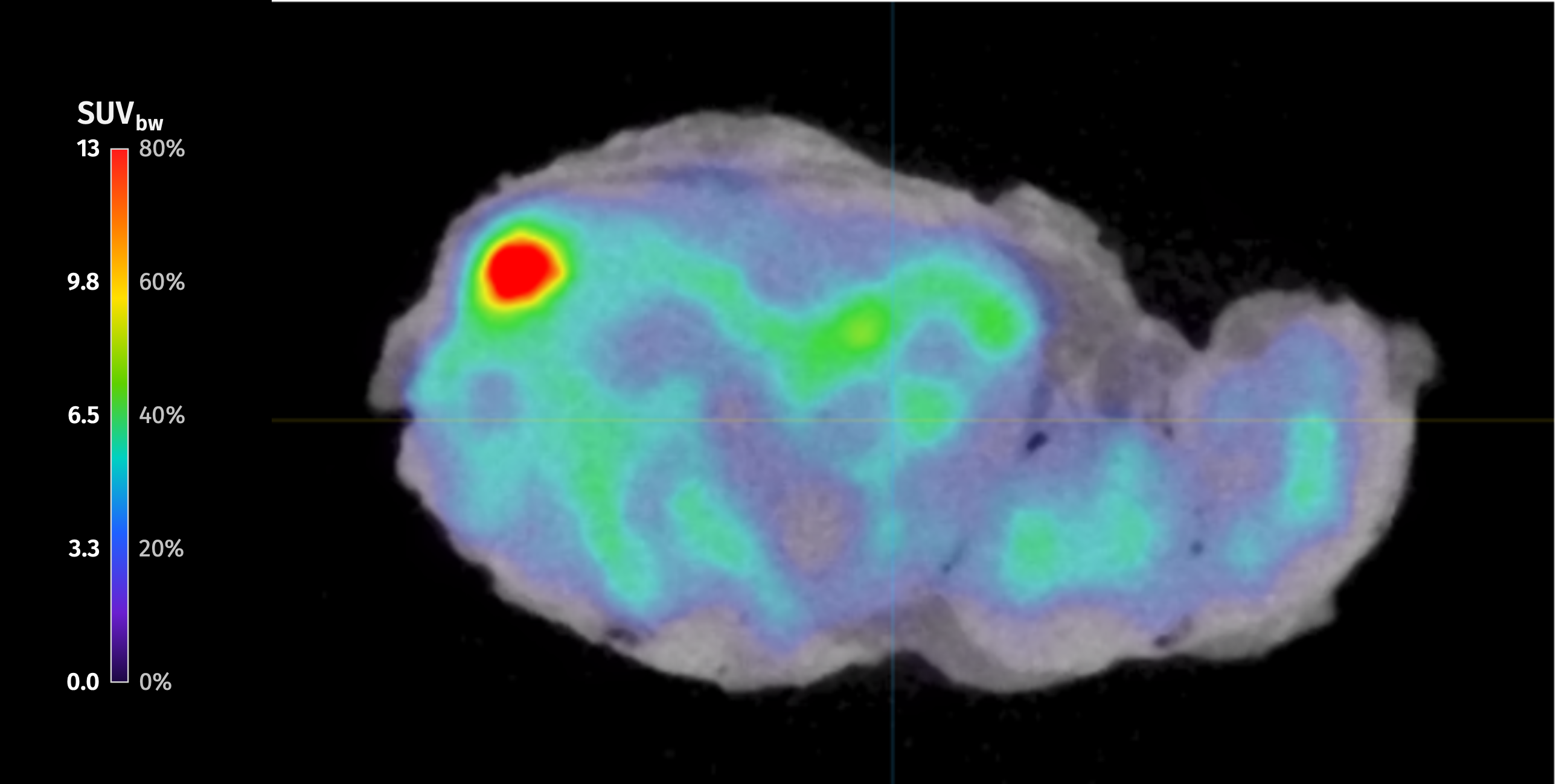
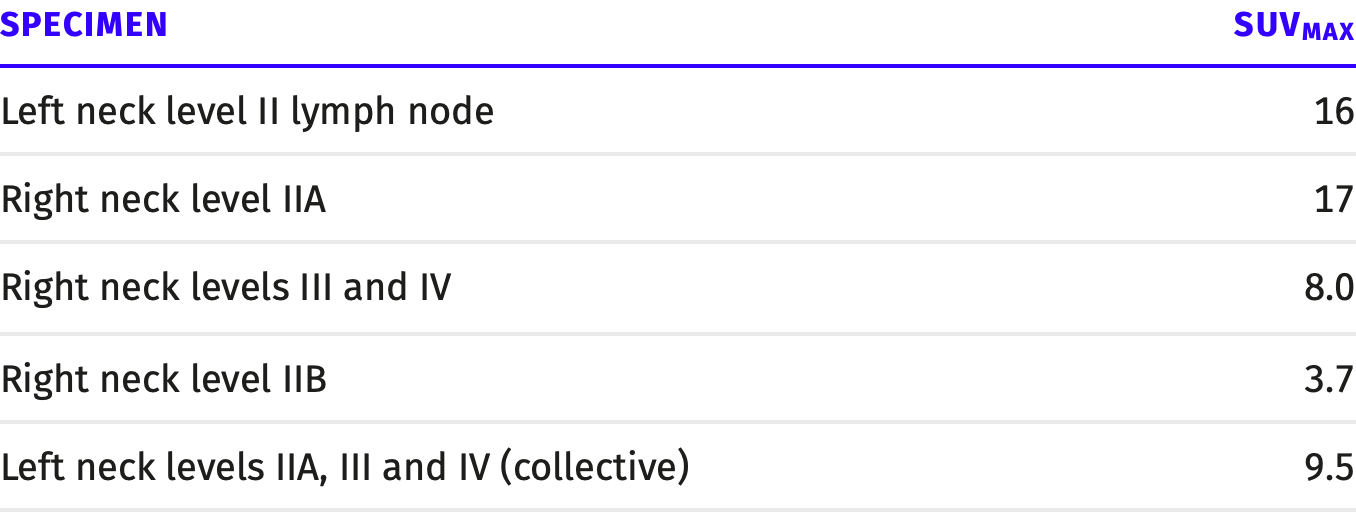
The contralateral left level II lymph node was excised, imaged with specimen PET-CT during surgery which suggested metastatic disease (SUVmax = 16). Based on this finding, bilateral level II–IV neck dissections were performed. Frozen section sampling confirmed the involvement of the left level II lymph node with squamous cell carcinoma, validating the decision to extend the left neck dissection.
Figure 1: High resolution PET-CT image of the contralateral left neck level II lymph node, which was confirmed on histopathology as metastatically involved. SUVmax (BW) = 16.Specimen PET-CT Imaging
The patient received an intraoperative injection of 0.8 MBq/kg (0.022 mCi/kg) of 18F-FDG per study protocol. During surgery, the left lymph node and nodal packets were scanned with the AURA 10 specimen PET-CT immediately after resection.
The contralateral left level II lymph node showed marked tracer uptake on specimen PET-CT, with an SUVmax of 16. This node had been labelled as indeterminate on preoperative PET-CT. Frozen section analysis confirmed metastatic squamous cell carcinoma.
The patient received an intraoperative injection of 0.8 MBq/kg (0.022 mCi/kg) of 18F-FDG per study protocol. During surgery, the left lymph node and nodal packets were scanned with the AURA 10 specimen PET-CT immediately after resection.
The contralateral left level II lymph node showed marked tracer uptake on specimen PET-CT, with an SUVmax of 16. This node had been labelled as indeterminate on preoperative PET-CT. Frozen section analysis confirmed metastatic squamous cell carcinoma.
Histopathological Evaluation
Frozen section analysis of the left level II lymph node during surgery was positive for squamous cell carcinoma. Final histopathology confirmed metastatic nonkeratinizing squamous cell carcinoma in 1 of 3 lymph nodes in this specimen, measuring 2.0 cm, without extranodal extension.
Results for the neck dissection specimens:
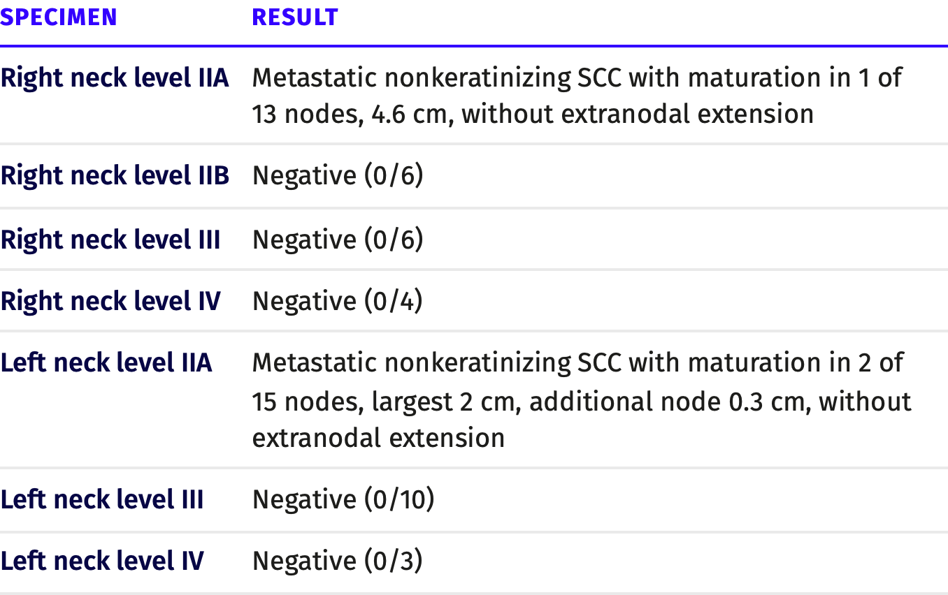
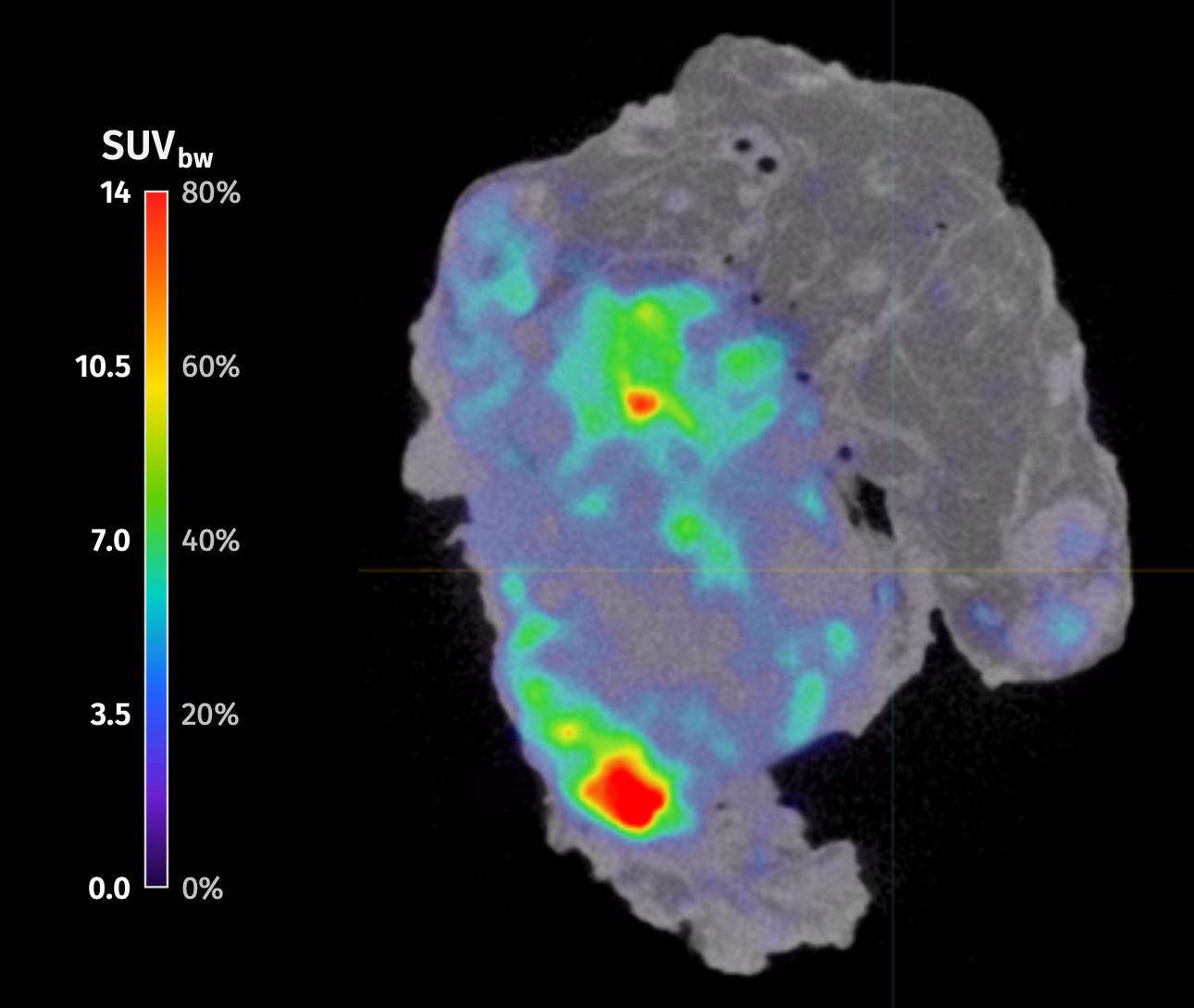 Figure 2: High resolution PET-CT image of the right neck level IIA lymph node package, which was confirmed on histopathology as metastatically involved. SUVmax (BW) = 17.
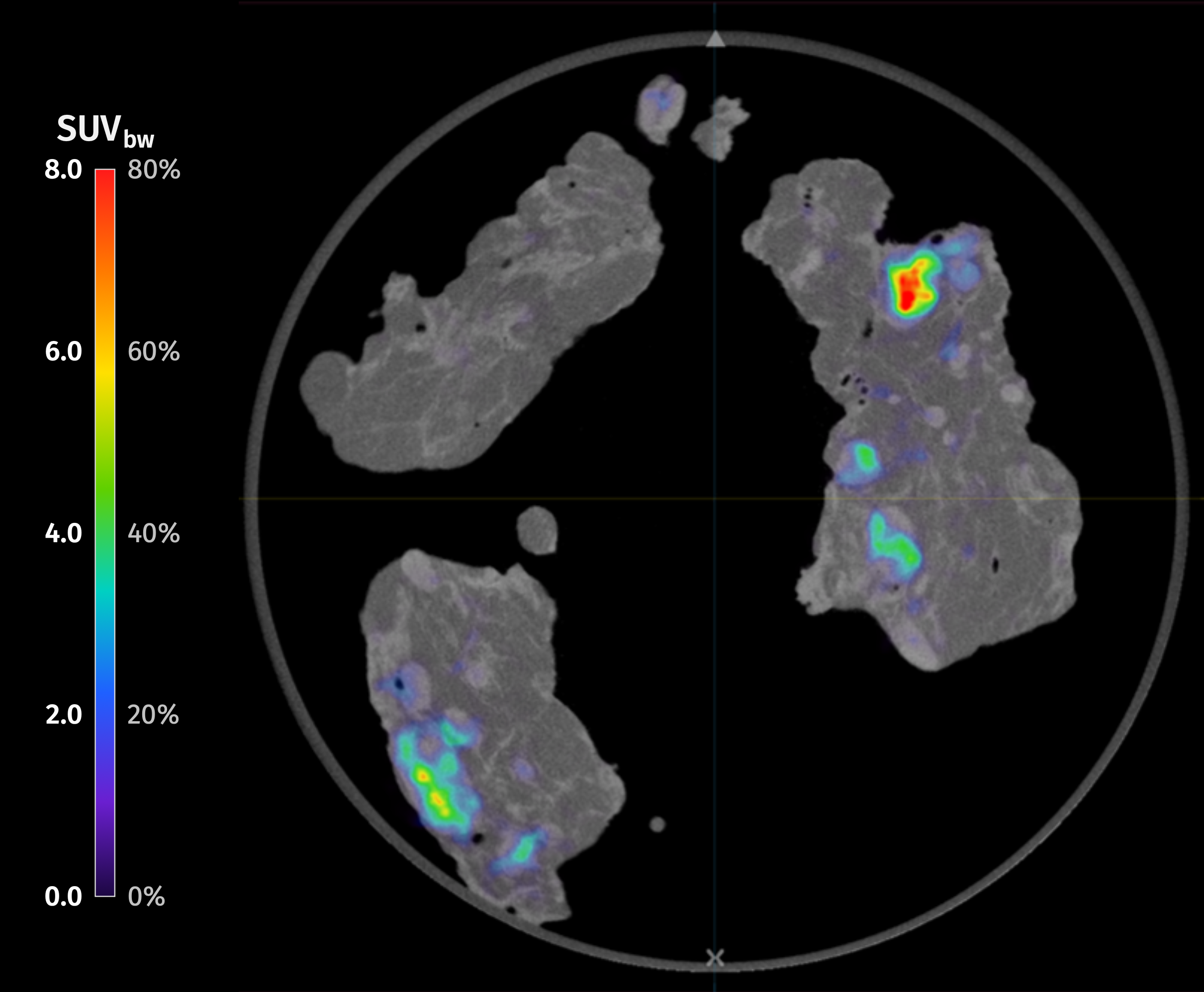 Figure 3: High resolution PET-CT image of the left neck level IV lymph node package (left upper quadrant), left neck level III lymph node package (left lower quadrant), left neck level IIA lymph node package (right). The first two lymph node packages were negative for SCC, while the latter was confirmed on histopathology as metastatically involved. SUVmax (BW) of the entire image = 9,5.
Discussion and Conclusion
This case shows specimen PET-CT imaging in a patient with HPV-associated squamous cell carcinoma of unknown primary origin. Preoperative PET-CT identified an indeterminate 1.2 cm left level II lymph node in the contralateral neck.
Specimen PET-CT imaging of the excised node during surgery showed marked FDG uptake (SUVmax = 16). Based on this finding, bilateral level II–IV neck dissections were performed. Frozen section analysis and final histopathology confirmed metastatic squamous cell carcinoma of 2.0 cm in the left level II lymph node. It also showed an additional 3 mm metastasis in left neck level IIA, while left levels III and IV were negative. The known right-sided nodal disease was also confirmed, with a 4.6 cm metastasis in right neck level IIA (SUVmax = 17 on specimen PET-CT).
This case highlights the potential of specimen PET-CT imaging during surgery to assess lymph nodes that are indeterminate on preoperative imaging. It can provide immediate information on nodal disease that supports decisions on the extent of neck dissection.
References
- Maghami E, Ismaila N, Alvarez A, Chernock R, Duvvuri U, Geiger J, Gross N, Haughey B, Paul D, Rodriguez C, Sher D, Stambuk HE, Waldron J, Witek M, Caudell J. Diagnosis and management of squamous cell carcinoma of unknown primary in the head and neck: ASCO guideline. J Clin Oncol.2020;38(22):2570–2596. doi:10.1200/JCO.20.00275
- Debacker JM, Schelfhout V, Brochez L, Creytens D, D’Asseler Y, Deron P, Keereman V, Van de Vijver K, Vanhove C, Huvenne W. High-resolution 18F-FDG PET/CT for assessing three-dimensional intraoperative margins status in malignancies of the head and neck, a proof-of-concept. J Clin Med.2021;10(16):3737. doi:10.3390/jcm10163737
- Walton GM, Adesanya O, Rowley L, Kallioinen SE, Torres-Rendon A, Matthews P, Agrawal R, Pickles C, Abdelrahman A, Naredla P. The use of intraoperative PET-CT in the assessment of surgical margins for patients with oral squamous cell carcinoma. Head Neck. Published online September 18, 2026. doi:10.1002/hed.70474